
Rudists: A fossil story

Jack Shimon (USA)
This article is adapted from a presentation given at the Denver Gem Show, September 17, 2016 by me, Jack Shimon. When I was six and a half years old, my Grandpa took me fossil hunting in central Texas. We went to a Carboniferous Limestone quarry that he had visited earlier and was given permission to enter and collect from. This was one of my first fossil hunting trips and I really enjoyed it. The ancient reef we went to (now a quarry) had huge boulders of limestone and tube-like things in it we later to be found to be rudist bivalves. This article is all about these finds and the efforts we went to, to find out what they were.


Fossils
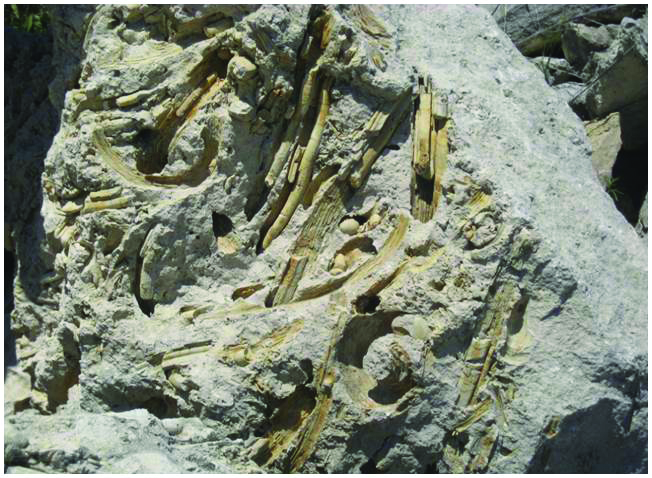
We spent a lot of time at the quarry observing the massive specimens onsite and then collected some smaller pieces to bring home and look at closer. A simple way of thinking about fossils is to consider them either as a cast or a mould. A mould is formed when an object is placed into a soft substrate and then decomposes or is washed away leaving an impression. This impression forms a cast fossil, when the mould fossil fills with sediment. I think cast fossils of these organisms were also forming when the hollow interior part of the animal was filled up (that is, inside their empty shells). We were curious about the mould fossil in Fig. 3 and what caused the spiny ridges lining it. The casts in Fig. 4 have a tube shape suggesting they might have grown as stalks. Looking closely, you can also see junctions in the stalk.


Using clues to solve the mystery
We were able to identify some fossils from the site, such as the ammonites in Figs. 8 and 9. These gave us a fossil age and also confirmed they came from a shallow marine environment. The ammonites were found shortly before we left when I was climbing on huge piles of rock and I was able to put pieces together to make a complete ammonite (Fig. 9).


We knew what our fossils were NOT, but still didn’t have an answer about what they actually are. So it was time to turn to the professionals. Since we were near the University of Texas in Austin, we started there. The university has a neat museum and we spent hours asking questions about our fossils, but no one knew what they were. It also has fossil drawers where kids are encouraged to match their local fossil finds to those from the Austin area, but none matched ours.
We continued our research online and came across a reef building organism called a rudist bivalve.
When I got back from my Texas trip, we still didn’t know for sure what we had found, so we took them to the Rock Fair at the Western Museum of Mining and Industry. I met a lot of exhibitors really interested in my fossils, but no one that could confirm they were rudists. Our last stop was to the Denver Museum of Nature and Science, where I was lucky to meet Dr Logan Ivy. He gave me a fascinating tour of the collections and spent time with me talking about my fossils. He positively identified them as rudist bivalves. One thing he explained that I had been wrong about was the function of the hook shaped valve. That part is not an anchor, although they do anchor to the sea floor with a small valve on the lower portion of the body. The hook-shaped valve is actually the top lid in this species.
Bivalves

of Zoology.)
After figuring out what our fossils were, we needed to know more about bivalves themselves. Bivalves are aquatic molluscs with a hinged shell such as mussels, scallops and clams. I’d seen some of these animals on beach trips, I just didn’t know they were called bivalves. I especially like the image in Fig. 11 of modern ‘edible’ bivalves because it shows a lesser known stalked bivalve called a razor shell. Also pictured clockwise from the top left are cockles, muscles and a scallop.

Rudist bivalves are extinct aquatic molluscs that came in many forms. They lived in shallow marine environments and dominated the reefs throughout the Cretaceous until they became extinct in the Upper Cretaceous. We found fossils of Caprinula and Titanosarcolites. Today their fossils can be found in limestone rocks.
There are two primary shapes that rudists take (Fig. 9). Elevator rudists attach to the sea floor and have an upright stalk. In Fig. 10, the rudists are probably Radiolites, very similar to the caprinulids we found, except for the shape of the lid. Recliner rudists lie on the bottom of the sea (Fig. 11). We only found one example at the quarry, a titanosarcolite. It is possible these fossils are washed away from where they originally grew before death, because the fossils don’t appear to be anchored to the seafloor.


Rudist bivalve morphology
The four main components of a rudist are the lower conical valve (which served as the anchor), the tube-shaped body, the internal body cavity (which ran the length of the outer body) and the upper hooked valve that served as a lid. Both valves are hinged and often fall off when the animal dies, which is why we were finding them separately at the quarry.
Taking a detailed look inside the animal, I think I can finally match the live animal with the fossil remains. It took a lot of research to understand what was happening in the live rudist and what changed as it became a fossil. The living rudist had a central hollow body cavity with chambers that closed off as it grew. This forms the segments we saw. The living rudist also had hollow outer tubes, much smaller in diameter, making up the shell. These aided in supporting the shell. When the rudist became a fossil, all the hollow spaces were filled with sediment, creating a cast of the original animal. The spiny ridges I observed in the mould of the fossil are actually casts of the outer shell tubes. Some of the fossils create a really nice picture of this morphology like the photo in Fig. 17.

Eureka! Mystery solved
You can see that the mould in Fig. 17 is lined with spiny tubes. These are casts of the outer body tubes. The central body cavity fits within this mould and was broken at several chamber junctions. The associated hooked upper valve is quite possibly embedded in the rock just below. It is amazing how a day digging at the quarry turned into an unexpected adventure.
References
Texas Water Development Board: http://www.twdb.texas.gov/groundwater/aquifer/GAT/.
The Rudists: http://www.ucmp.berkeley.edu/taxa/inverts/mollusca/rudists.php.
Wooster Geologists https://woostergeologists.scotblogs.wooster.edu/2011/01/23/wooster%E2%80%99s-fossil-of-the-week-a-most-unlikely-clam-rudists-from-the-upper-cretaceous-of-the-oman-mountains/.
Bureau of Economic Geology, the University of Texas at Austin. Stuart City Trend, Lower Cretaceous South Texas.
Fossils Explained: Rudists: http://www.academia.edu/1316778/Fossils_Explained_Rudists.