
Phenomenal fossil fern: Forgotten for 40 years

Stephen McLoughlin (Sweden), Benjamin Bomfleur (Sweden) and Vivi Vajda (Sweden)
On some occasions, it is the hard sweat and toil of palaeontologists labouring in the field at carefully planned excavation sites that yields the prize specimen on which careers are built. On other occasions, it is the chance discovery by an amateur collector that may yield that special fossil. We present an account of one such remarkable fossil discovery by an eccentric farmer in southern Sweden. However, more remarkable is that this exceptional fossil remained unstudied and largely unnoticed in a major museum for almost 40 years, before its true significance was realised.
The story begins near Lake Korsaröd, in the heart of the southern Swedish province of Scania. Gustav Andersson (born 16 May 1915; Fig. 1) owned a small homestead bordering the shores of this lake.

Although Gustav made a living from farming, his true passion was natural history and he even adorned the walls of his house with his own sketches of Mesozoic scenes. Although he never received any formal scientific training, Gustav was an avid reader and had a keen eye for nature. He used these skills to identify a great range of plants on his property down to the rare ground orchids that episodically bloomed on the local volcanic soils. He also identified Neolithic burial sites, flints, stone clubs and other ancient human artefacts that lay scattered about the landscape.
However, his particular interest was geology. Gustav identified several Jurassic volcanic plugs and Quaternary glacial moraines in the Korsaröd district that had not been recognised by the professional geologists of his day. He was a keen observer of the subtleties of the landscape, which had been shaped by volcanism, rivers and the great Quaternary ice sheets that had covered Scandinavia. He often wrote to the geology professors at Lund University to announce his ideas. Often dismissive of his claims, the academic geologists sometimes had to eat their words, when Gustav patiently took them into the field to show them his discoveries.
Perhaps Gustav’s most important discovery came in the late 1960s or early 1970s. On that occasion, he identified a volcanic ash bed, containing a fossil log near the shores of Lake Korsaröd (Fig. 2).

At that time, some geologists argued that the volcanic rocks of central Scania were of Cenozoic age, but Gustav argued that his discovery of an entombed petrified log pushed the age of volcanism back to the Mesozoic. He sent off a letter to Lund University to report his discovery. The Lund geologists, ever wary of his theories but nonetheless acknowledging his keen observational skills, dispatched two young men to excavate the site in search of more fossil wood. Unfortunately, Gustav was away at the time, but the two young men laboured for a day and found nothing of value. Some days later, a letter arrived from Professor Helmquist in Lund stating that the site contained nothing of interest for geologists.
Gustav was not impressed. Taking his pick and shovel, he dug deeper into the trench and soon found several pieces of wood up to 7kg in weight. These samples (Fig. 3) he sent to Hans Tralau, who was then an assistant curator at the Swedish Museum of Natural History in Stockholm. Tralau studied the fossil spores and pollen entombed in the ash deposit that contained the wood and confirmed that the beds were indeed of Early Jurassic age.

Among the many samples of volcanic rock and fossil wood that Gustav Andersson sent to Hans Tralau was a single peculiar petrified fern rhizome (Fig. 4).

Tralau clearly recognised the significance of this fossil because he prepared several thin sections and took a series of photographs of the specimen (Fig. 5). Undoubtedly, Tralau intended to publish a formal description of this fossil, but his untimely death in March 1977 meant that the project was never completed. Archival correspondence at the Swedish Museum of Natural History reveals that, some years later, Britta Lundblad, then head of the Palaeobotany Department, exchanged letters with Gustav Anderssson, also indicating her intention of writing a report on the material. However, her retirement in 1986 left the task unfinished. After that, the fossil lay in the collections and largely went unnoticed by the curatorial staff or the large number of visiting researchers.

In 2013, we opened the drawers and looked again at this curious fossil. It is a small specimen – just 7cm long and 4cm in diameter – but good things come in small packages. The specimen is a three dimensionally preserved fern rhizome, clothed in a mantle of frond bases. It belongs to a distinctive family of ferns – the Osmundacae or Royal Ferns. This family has a very impressive fossil record extending back to the Permian and was distributed globally, but the Korasröd fossil is the only permineralized (three-dimensionally preserved) example yet known from the Mesozoic of Europe – a character that originally attracted our attention. However, this is not the most remarkable aspect of the fossil. Its truly unique qualities are only apparent when thin sections of the stem are studied with a transmitted light microscope.
In most permineralized plants, it is only the thick walls of the water-conducting (xylem) or periderm (bark) cells that are preserved, since these are robust structures composed of lignin or suberin that are resistant to decay. This gives sufficient time for silica-, calcium-carbonate- or other mineral-bearing fluids to percolate through the pore spaces in the tissue, precipitate minerals on the cell walls and infill cell cavities. Softer food-conducting (phloem), pith and cortex tissues generally decay before the permineralization process is complete. In a few special cases, such as the Rhynie Chert in Scotland, the Grand-Croix cherts in France and the Princeton Chert in Canada, even some thin-walled cells of delicate stem and leaf tissues are preserved.
The preservation of Gustav Andersson’s fern from Scania goes one or two steps further. Careful examination of the stem tissues in thin section revealed that even the most delicate pith cells are preserved (Fig. 6).

Moreover, in the centre of each cell is a distinctive brown-stained nucleus (Fig. 7).

At higher magnifications, it is even possible to discern a minute nucleolus preserved in the centre of most cell nuclei. Even more exceptional is the fact that a few cells appear to have been preserved at various stages in the process of cell division (mitosis) (Fig. 8). These cell nuclei show successive steps in the breakdown of the nuclear membrane, unravelling of the coiled chromosomes and apparent alignment of chromosomes in the lead up to cell division. A few cells around the periphery of the stem also show nuclei in which the nuclear contents have contracted to a distinctive crescent shape. This is a feature typical of programmed cell death (apoptosis).

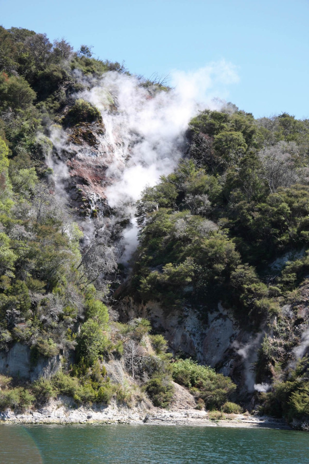
The preservation of these remarkably transient features in the life cycles of cells gives us a clue as to the speed of the permineralization process. Studies of modern hot spring environments, such as Yellowstone National Park, have revealed that plants can become encrusted with mineral coatings within days to weeks. However, since some of the stages of cell division that we see in the Korsaröd fern last no more than about 15 minutes in living plants, we interpret this Jurassic fern to have been permeated by mineral-rich fluids causing at least fixation of intracellular processes on a time scale of only minutes to hours.
Using light microscopy, various other very fine granular contents are evident within the cells, but they are difficult to identify. Scanning electron microscopy (SEM) gives us greater power to resolve these minute structures. We etched the polished surface of the fern in dilute acid and, with the aid of SEM, we were also able to resolve fine details of pitting on the water-conducting cells (Fig. 9) and clusters of spherical bodies in some pith cells that appear to be amyloplasts – the organelles responsible for the synthesis and storage of starch granules.

Such spectacular preservation opens up a whole new field of research – palaeocytology – the study of cell contents in deep time. There have been a few other cases of exceptional preservation in plants, algae and early animal embryos, in which cell nuclei or cell division have been reported. Many of these cases remain controversial, but a comparison between the cells of the Korsaröd fern and the cells of its modern relatives (Osmunda regalis and Osmundastrum cinnamomeum) leaves no doubt that true nuclei are preserved in the Jurassic fossil.
We suspect that preservation of cellular contents is more common that generally assumed. In recent years, convincing evidence has been forthcoming for the preservation of osteocytes (bone-producing cells) with nuclear material in Cretaceous dinosaur bones, colour pigments in fossil bird feathers and chloroplasts in Eocene conifer leaves. With advances in analytical techniques, especially synchrotron X-ray tomographic microscopy (Fig. 10), we predict that many more examples of fossil cell organelles will be revealed in the near future.

Finding fossilised cell nuclei is surprising and exciting, but what can it really tell us about the plants other than the speed of their preservation? After all, it is not that we didn’t expect Jurassic plants to have nuclei in their cells. The Royal Ferns (Fig. 11) have long been considered an archaic group within the main lineage of ferns.

They are generally considered to represent the descendants of the first branch that diverged (in the Permian) from the ‘leptosporangiate’ fern evolutionary tree. Certain members of the Royal Fern family are considered to be very conservative in an evolutionary sense and have sometimes been dubbed ʻliving fossilsʼ. That is, some show morphological and anatomical characters that have not changed significantly since the Mesozoic. Some fossils are considered so similar to their living relatives that they have been assigned to the same species.
The most extreme cases of this are fossils recorded from the Late Cretaceous of North America that have been assigned to the living species Osmundastrum cinnamomum; and Osmunda claytoniites from the Triassic of Antarctica, which is considered almost indistinguishable from the modern species, Osmunda claytoniana. Although these morphological similarities have been assumed to result from genetic conservatism, thus far we have lacked fossil evidence to directly assess similarities between the genomes of modern and fossil species.
One of the important factors in plant evolution is polyploidy – the duplication of the number of chromosomes within the genome, which can occur spontaneously by processes such as meiotic or mitotic failures, and the fusion of unreduced (diploid) gametes. Polyploidy is much more common in plants than animals and some estimates suggest that 30% to 80% of modern plant species are polyploids. Around one-third of fern speciation events are interpreted to have involved duplication in the number of chromosomes.
All modern members of the Royal Ferns have a chromosome count of 22. Given that the size of cell nuclei is proportional to the quantity of genetic material that must be packed into the nuclear membrane, we were able to compare the size of the nuclei in the Jurassic fern to nuclear sizes of their modern relatives to determine if any ploidization events had taken place in this group over the past 180Ma. Our measurements confirm that the nuclear sizes of cells in the Korsaröd fossil and in modern Osmundastrum cinnamomeum match very closely. This confirms that the genome size of the Royal Ferns has indeed remained essentially unchanged since the Early Jurassic — a remarkable example of evolutionary stasis.
The fossil also has a role to play in the way we classify Royal Ferns. Recently, genetic evidence has been used to segregate one species that was traditionally placed in Osmunda into a new genus Osmundastrum. The Jurassic fossil has key anatomical characters that are shared by both taxa suggesting that there is no clear-cut distinction between these genera.
Returning to the matter of the remarkable quality and speed of cellular preservation, one must ask: in what sedimentary environment could this occur? We interpret the host rock at Korsaröd to be a lahar (volcanic debris flow) deposit associated with the eruption of a small volcano in central Scania during the late Early Jurassic. We suggest that the fern rhizome, associated conifer woods and other plant debris were caught up in the debris flow and that hydrothermal fluids were rapidly flushed through the sedimentary system in association with the volcanism (Fig. 12). These supersaturated fluids permeated the plant material and precipitated calcium-carbonate in and around the cells before any significant decay had occurred – indeed while most cells still retained their cytoplasmic contents.
Finally, if this model for the permineralization of cellular details is correct, then it also has significance for our search for evidence of ancient life on Mars. Hydrothermal systems represent particularly favourable conditions for both the survival of life (supplying geothermal energy, water and a concentrated mineral supply) and its fossilisation (through rapid mineral precipitation). Swift burial of organic matter by debris flows potentially enhances these chances of preservation. Since volcanism and associated debris flows, together with hydrothermal activity, were apparently common on Mars in its early history, such deposits are likely to be high on the priority list of targets in the search for Martian fossils by the current NASA robot rovers, Opportunity and Curiosity.
Sadly, Gustav Andersson died on 27 May 2002, so he did not live to see the full significance of his fossil brought to light. Nevertheless, the enthusiasm, astute observations and avid collector’s mindset of amateur fossil hunters like him continue to make important contributions to the cutting edge of palaeontology around the world.
Further reading
Bomfleur, B., McLoughlin, S. & Vajda, V., 2014. Fossilized nuclei and chromosomes reveal 180 million years of genomic stasis in Royal Ferns. Science 343, 1376–1377.
About the authors
Stephen McLoughlin and Benjamin Bomfleur both work at the Swedish Museum of Natural History, Stockholm (steve.mcloughlin@nrm.se; benjamin.bomfleur@nrm.se), whereas Vivi Vajda worksat the Department of Geology, Lund University, Lund (Vivi.Vajda@geol.lu.se).
Stephen McLoughlin was raised on a farm in Queensland, Australia, where his first introduction to fossils was collecting Jurassic petrified wood turned over by the plough. He is currently a senior curator of palaeobotany at the Swedish Museum of Natural History, Stockholm. He works mainly on the systematics, evolution and palaeoecology of late Palaeozoic and Mesozoic seed-plants, ferns and lycophytes. He is presently studying fossil plants from Antarctica, Sweden, Australia and China. He previously worked as a lecturer and researcher in several Australian universities and has carried out fieldwork on all continents.
Benni Bomfleur grew up in northern Germany and is a researcher at the Swedish Museum of Natural History, Stockholm, where he is currently studying the evolution of the Royal Fern family, the anatomy of Permian seeds from Australia, and the palaeoecology of Antarctic Mesozoic plants. Benni has previously been a postdoctoral researcher at the University of Kansas. He is a veteran of several palaeontological fieldtrips to remote regions including the Transantarctic Mountains, Ellesmere Island in Canada, and the Nullabor Plain in Australia.
Vivi Vajda grew up in southern Sweden and is now a professor of paleontology at Lund University. Her research focuses on fossil spore and pollen assemblages, which she uses to determine the relative ages of sedimentary rocks, and particularly the rate of vegetation change through major events in Earth’s history. Plants respond rapidly to climate changes and to catastrophes such as asteroid impacts and large volcanic eruption phases. Vivi’s investigations focus on Mesozoic sediments from Sweden, New Zealand, China, and Denmark. She is also active in international geo-related organizations being former chairs for both the UNESCO geosciences program IGCP and the Swedish Geological Society.